INDEC BioSystems
Multiple Solutions in
a Single Package
|
|
|
| -
Image Snap and Archive |
-
Time Lapse |
|
|
- Fluorescence |
-
Multichannel |
|
Imaging Workbench 6 is a program that
acquires and analyzes images that change over time. It is most often
used for low light level fluorescence applications in biology.
During acquisition, Imaging Workbench controls wavelength switching
devices and cameras to acquire precisely timed series of images and
save them to disk.
During on-line and off-line analysis, Imaging Workbench calculates
average ratio, ion concentration or intensity over outlined regions.
The data are exported to spreadsheet program-readable files.
Imaging Workbench performs with great stability and tight timing.
It is compatible with Windows 10. Armed with multiple-window
display, fast acquisition and wavelength control, Imaging Workbench
6 is ideal for measuring the dynamic process of ion concentration
changes inside cells. In addition, Imaging Workbench 6 runs concurrently
with Axon Instruments' Clampex 8 to 10, performing simultaneous acquisition
of electrophysiology data. Cooperative electrophysiology-imaging data
review can be performed during analysis.
Key Features
Image Acquisition
- Acquire images in ratiometric and nonratiometric modes
- Acquire images using one or two dye-sets. Either can be ratiometric
or nonratiometric

- Simultaneous control of excitation and emission wavelength for
each dye-set
- Live video mode for focusing
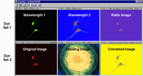
- Shading correction (see image below)
- ΔF/F0 for any nonratiometric wavelengths; F0
can be averaged to a low-noise image
- Background subtraction
- View and save a smaller acquisition area in the field of view
to accelerate acquisition and save hard disk space
- Store images to a ring buffer for the highest performance
- Always-on-top Acquisition Control Panel for on-the-fly changes
to acquisition parameters
Image Display

- Generates ratio images, pseudocolor mapped based on ratio or intensity
values
- Annotates image files with comment tags and voice tags
- Prints on-line graphs. Copy and paste graphs into other programs
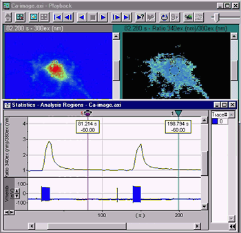
- Dragging a "sync" cursor along the time-axis of the graph will
automatically display the image acquired at the corresponding time
(see below)
Timing Control
- Switch acquisition rates at will when recording continuously
- Allows episodic acquisition protocols, e.g. with electrophysiology
- Precise timing control of camera, wavelength changers and shutter
devices
- Timing Overview window that illustrates device control and image
acquisition timing, allowing for precise calculation of fluorescence
exposure cycles

Concurrent Imaging and Electrophysiology
Imaging Workbench 6 has enhanced features for cooperative, simultaneous
imaging and electrophysiology when used in conjunction with 8 through
10. IW is also compatible with PatchMaster v2 from Heka.
- Runs concurrently with Clampex or PatchMaster
- Receives software trigger from Clampex to start image acquisition
- Digital outputs are sent through the INDEC BioSystems DIO-3 interface box
or equivalent. Digital inputs to the DIO-3 can trigger image acquisition
- Displays electrophysiology data, fluorescence intensities and/or
ion concentrations in the same graph
- A "sync" cursor can be placed anywhere on the electrophysiology
data, and the image acquired at the corresponding time will be automatically
displayed

- Automatically opens the most recently acquired Clampex data file
at the end of image data acquisition
Image Analysis
- On-line intensity and ratio or concentration calculation
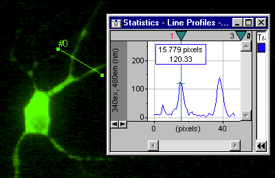
- Line profile

- Post acquisition analysis
- ΔF/F0: average F0 over images for lower noise
- 16-bit multi-image TIFF export
Computer System Configuration
for Imaging Workbench
Recommended:
- Operating system: Microsoft Windows 10
- System and CPU: IBM-PC compatible with Pentium IV CPU 2+ GHz
- Memory: 4 GB depending on activity
- Hard disk: 80 to 200 GB hard disk depending on activity
- Graphics card: 24- or 32-bit color on a 1280 x 1024 pixel monitor
with 70 Hz refresh rate
- Monitor: compatible with the graphics card specifications; two
monitors can be helpful
Device Control
- PCI slot for camera interface board or video digitizer
- PCI slot if using wavelength switcher from TILL Photonics or Cairn
Research
- Parallel port if using Sutter Instrument wavelength switcher,
Uniblitz shutter and/or digital I/O
- Extra parallel port if using pCLAMP in addition to parallel-port
device control
- Serial port if using serial port device control
- USB port
Minimum:
- Operating system: Microsoft Windows 10
- System and CPU: IBM-PC compatible with Pentium processor
- CPU speed: 1GHz for Windows 10
- Memory: 4GB for Windows 10
- Hard disk: 80 GB depending on activity
- Graphics card: 16-bit color on a 1024 x 768 pixel display
- Monitor: compatible with the graphics card specifications
Device Control
- PCI slot for camera interface board or video digitizer
- PCI slot if using wavelength switcher from TILL Photonics
- PCI slot if using wavelength switcher from Cairn Research
- Parallel port if using Sutter Instrument wavelength switcher,
Uniblitz shutter and/or digital I/O
- Extra parallel port if using pCLAMP in addition to parallel-port
device control
- Serial port if using serial port device control
- USB port
Hardware Support
Supported Digital Cameras
- Andor iXon and luca series
- Hamamatsu ORCA series
- Photometrics cameras from Roper Scientific, including Cascade,
CoolSNAP, Quantix and SenSys
- SensiCam VGA, SVGA, QE and EM, and Pixelfly series, from PCO
Computer Optics / The Cooke Corp
- Selected Princeton cameras from Roper Scientific
Supported Analog (Video) Frame Grabbers
- Data Translation DT3155 frame grabber for PCI bus
Supported Illumination Control Devices
- Sutter Instrument Lambda 10, 10-2, 10-3 and 10-B filter wheels
- Sutter Instrument Lambda DG-4 and 5 wavelength switcher
- TILL Photonics Polychrome II, IV and V monochromator
- Cairn Rotor filter wheel and Optoscan monochromator
- Cairn OptoLED multi-LED light source
- Other wavelength switchers may be supported through script files
See more supported hardware
