Integrated Systems
for Cellular Imaging

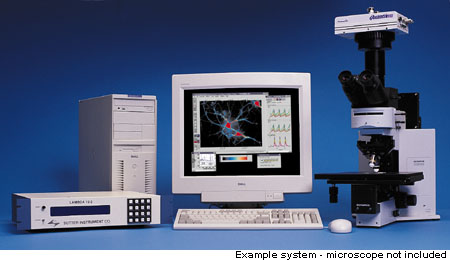
The Integrated Imaging System is
used by cell biology and physiology researchers to determine ion concentration,
dynamic fluorescence, intrinsic signal and cell volume activity in
live preparations using fluorescence and/or bright-field microscopy.
The Integrated Imaging System is essential for concurrent imaging and
electrophysiology or imaging and photometry.
Imaging Workbench provides these custom systems because scientists
have been asking for complete systems in which:
- They can choose the best imaging components for their budget and
applications, with the help of an experienced specialist;
- The functions they want can be performed simply and elegantly;
and
- They are able to test each of the components in their lab and
verify acceptable performance.
Users can choose the best components available for biological imaging.
The system is then demonstrated in your laboratory so you can verify
the performance.
We are continually adapting the system to the new demands of researchers.
The migration pathways we are following include support for high-speed
imaging (taking advantage of new fast cameras) and combining electrophysiology
and imaging.
